Prologue
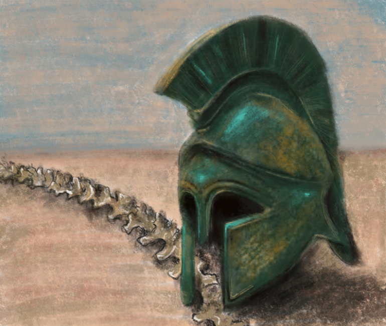
Have you heard the story of Homer’s Achilles? Once hailed as the greatest mythical warrior in all of Ancient Greece, he now serves as a cautionary tale to protect one’s vulnerabilities. Achilles’ vulnerability was his heel–the only part of his body not granted invincibility at birth. Similar to Achilles’ heel, we too are born with a part of our body so crucial to our being that it cannot be repaired or replaced: the central nervous system (CNS).
Composed of both the brain and spinal cord, the CNS synthesizes sensory information collected throughout the body by coordinating conscious and unconscious reactions with the brain. For instance, on a bright day, your brain might receive a message through the spinal cord that the sun is hurting your eyes. In response, your brain would send a signal through the spinal cord to direct your hands to shield your eyes from the sun. The CNS is a sophisticated system and is critical in processing and generating responses to external stimuli, therefore any mutations or external damage to it could significantly alter a person’s quality of life.
Within the robust neuroscience research hub at UC San Diego, the Gleeson and Zheng Labs analyze cellular mechanisms for CNS deterioration at opposite ends of the age spectrum. Headed by Dr. Joseph Gleeson, the Gleeson Lab studies disease mechanisms impacting CNS development. This lab has begun to uncover how genetic anomalies result in neural tube defects within the fetuses of mothers who are low in folic acid. On the other hand, the Zheng Lab headed by Dr. Binhai Zheng studies the extent of the spinal cord’s regenerative abilities, specifically the repair mechanisms within CNS neurons.
Act I: Abnormalities & The “Gene”rosity of Fate
In embryonic development, the CNS begins as a sheet of cells until around week five, when it folds into a tube. From there the caudal, or lower end of the tube, extends to form the spinal cord while on the opposite side, the cranial end, enlarges to form the brain. Developments within the embryo can make it challenging for the sheet of cells to fold properly. In some cases, the tube doesn’t fully close by the end of week four, forcing it to stay open throughout the remainder of development. Conditions where the spinal cord fails to close properly are known as neural tube defects (NTD). NTDs are amongst the most common CNS structural abnormalities, currently affecting around 5% of all children globally.
Within this percentage, the Gleeson Lab is focusing their research on a specific type of NTD called spina bifida. Spina bifida is a congenital neurological disorder in which the spinal cord does not close properly, inhibiting the formation of the backbone. In addition to leaving the spinal cord more exposed to extraneous dangers (i.e. accidents), spina bifida also limits the brain’s range of communication with the body. For instance, children with the most severe form of spina bifida, myelomeningocele, are born with a small pouch on their back containing part of the spinal cord, which usually limits walking and sensory capabilities.
On his mission to treat these afflicted children, Dr. Gleeson is partnering with the Spina Bifida Association (SBA) to explore the genetic and environmental implications of the condition. Years of research in the field of neuroscience have shown a pattern, although still unclear, of NTD inheritance and a correlation between low levels of vitamin B9, or folic acid, and risk of developing a NTD. This suggests that there are multiple factors that lead to a fetus developing a NTD. Therefore, Gleeson and his team believe that the key to understanding spina bifida lies in examining not only the child, but the parents as well. Using trio exome sequencing, or the process of extracting and sequencing a child’s and parents DNA, the Gleeson Lab can compare their genes against one another and identify the specific ones responsible for causing the condition. Ultimately Dr. Gleeson and his team aim to improve understanding of the genes responsible for spina bifida and develop treatments, particularly for patients with less developed spinal cords.
Act II: Resurrecting Dead Cells through Genetic Discovery
As cells age, their ability to repair their injuries greatly reduces. Spontaneous self-repair distinguishes the axons in the central nervous system (CNS) from the peripheral nervous system (PNS): PNS axons can readily regenerate, while CNS axons cannot. Axons, or components of nerve cells called neurons, act as a channel for electrochemical signals to relay sensory information to the brain and carry motor output to various parts of the body. As the central communicators to and from the brain, CNS axons that facilitate interpretation of stimuli from our environment are especially essential for survival.
With this in mind, the Zheng Lab studies the molecular mechanisms behind axonal repair in the CNS and how its induction can treat patients with permanent motor function loss. Two of the Zheng Lab’s graduate student researchers, Camilo Londoño and Kween Agba, explained how their lab began to explore these mechanisms by identifying genes that directly suppressed axon regeneration in the CNS.
In 2023, Dr. Hugo Kim attempted to regenerate axons within the corticospinal tract (CST) by silencing genes responsible for axon suppression within experimental mice. The CST was selected because of its integral role in providing voluntary movement; paralysis or other forms of limited motor function occur when axons in this path are damaged. Mice were injected in the middle of their spinal cord with one gene-specific retrograde viral tracer, which showed how information travelled to the brain through this pathway by glowing green under a microscope. This tracer also deleted genes in the CST believed to suppress general axon regeneration. After four weeks, researchers inflicted damage in the same spinal cord region and a red gene-specific retrograde viral tracer was injected to follow the same CST pathway. Presence of the red and green tracers throughout the CST pathway meant that the CST neurons still allowed information to travel to the brain despite their injuries. Dr. Kim saw that while many neurons fluoresced either green or red, a sizable sample overlapped. These cells, categorized by neuron type, location, and gene expression, were input into a new cell identification program called a Regeneration Classifier.
Agba and Londoño posit that this Regeneration Classifier program will be an integral tool for understanding the extent of CNS neuron regeneration. As more information is added to the program’s database, researchers can use the program to create a spatial map of the CNS to indicate which neuronal cell types in regions of the CNS have higher regenerative capabilities. Researchers and medical professionals could match these regions to areas commonly associated with permanent musculoskeletal impairment. Regenerative axon populations in the brain could further be tracked by experimenting with mice at different ages. By seeing how these populations grow or shrink as one ages, the long term efficacy of genes coding for axon suppression can be discovered as well. If regenerative axon growth maps are created using the Regeneration Classifier program, researchers can pinpoint which musculoskeletal diseases react with gene therapy and when in a person’s life gene-based spine treatments work best.
Epilogue
The spinal cord is a powerful tool that can be permanently damaged if mishandled or malformed. While Achilles never recovered from the wounds to his heel, thanks to the Gleeson and Zheng labs, we can use our understanding of the mechanisms behind neural tube growth and CNS axon regeneration to discover how to overcome defects and injuries to this organ.
With discoveries of the spinal cord formation and regeneration on a molecular level, the health policy and pharmaceutical landscape can fundamentally change. Treatments for neural tube defects and spinal cord diseases can relieve patients and caretakers of long-term healthcare costs like rehabilitation and specialized care. Patients and their families will also be relieved of the psychological burden stemming from financial and treatment stress. While some aspects of healthcare like spinal-cord-specific rehabilitation services may decline in use, knowing how these defects and injuries form can advance fields in spinal cord academia, as well as pharmacology and biotechnology by creating new medication and prosthetics. Perhaps someday, spinal cord repair and defect prevention could be such a commonplace procedure that future generations see its current vulnerabilities like we see Achilles: a myth.

