With the current COVID-19 pandemic, children experience less social interaction than possibly ever before. Ideally, they would continuously learn and socialize with classmates in classrooms, rather than through online video meetings. However, more children and people of all ages lack proper levels of social interaction, causing worry for how this unprecedented social isolation could impact individuals. How do lonely environments impact people on a biological level?
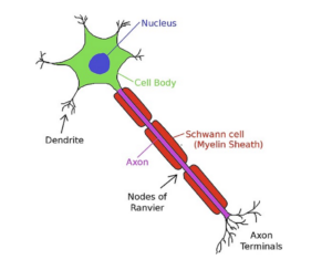
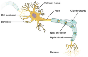
To understand how social isolation affects the body, we must first understand how the nervous system functions. Neuron cells consist of a cell body and a long axon that transmits impulses across the neuronal network, and myelination occurs when oligodendrocyte cells wrap neurons with myelin. Myelin is composed of layers of phospholipids and proteins that insulate nerve fibers and increase the speed of impulse transmission throughout these nerve fibers. Speeding up impulses highly benefits neuronal communication, or signaling between nerves throughout the body, and a lack of myelination can lead to cognitive decline. Some of the proteins which oligodendrocytes produce, in order to myelinate neurons, include MAG and MBP.
Scientists, Manabu Makinodan, Kenneth Rosen, Susumu Ito, and Gabriel Corfas experimented on young mice to gauge how social experience impacts the nervous system. While neuroscientists previously believed that neurons are the primary cells contributing to overall nervous system function, recent studies suggest that neuroglial cells, such as oligodendrocytes, are also highly significant in the nervous system due to their impact on proper neural function, inducing interest in the field of neuroscience. This is largely due to the discovery of multiple mechanisms through which neuroglial cells impact the excitation and inhibition of nerves.
The scientists’ experiments involved social experiment tests on mice, as well as administering the chemical agent, tamoxifen, to assess the amount of myelination under different conditions. The results demonstrate a correlation between less exposure to social interaction and less myelination, and the injection of tamoxifen leads to the same result. According to these results, there is a critical period during which young mice should be exposed to social experiences in order to maximize the myelination of their central nervous system.
One of the conducted experiments, an object displacement test, observed the mice’s interest in an object to test their working memory. The mice were initially exposed to a space in which a specific object would be placed, and four weeks later, they were placed in the same space, this time without the object present. The results show that mice that lived with other mice in a regular environment tended to have more interest in where the object had gone, while isolated mice showed less interest in the missing object. In addition to gauge the mices’ interest, the object displacement test also demonstrated a difference in the accuracy of their working memory. In this scenario, a higher percentage of correct responses refers to the amount of times the mice found the location of the object. Mice which were not isolated earned a higher percentage of correct responses when entering the space with the displaced object. Isolated mice, willing to look for the object, had a significantly lower percentage of correct responses than the mice in a regular environment, indicating that socialized mice displayed a better working memory.
In addition, further results from the same social tests on mice, observing interest and memory, show that the oligodendrocytes of regular mice have more intricate and complex morphologies (connection between biological structure and function) when compared to isolated mice. For instance, isolated mice have shorter oligodendrocyte extensions, resulting in less overall myelination, as oligodendrocyte extensions reach out to less neurons.
In their experiments, the scientists also studied the mices’ myelin. Firstly, they concluded that isolated mice showed reduced levels of the MBP and MAG proteins in comparison to regular mice. They also determined that isolated mice have a much higher G-ratio. The G-ratio is the ratio of the inner axonal diameter to the outer diameter, and indicates the amount of myelination that can occur. The inner axonal diameter reflects the axon’s diameter without myelination, and the outer axonal diameter marks the diameter of the axon with myelin. Less myelin leads to a smaller outer axonal diameter and a higher G-ratio, suggesting that isolated mice with higher G-ratios experience less myelination without change in axon diameter. This means that the extent to which myelination occurred became reduced with isolation, in addition to a reduction in the amount of myelinated neurons.
Despite the wealth of research linking social isolation to less myelination and neural communication, additional experiments show that the isolation of mice after a certain age does not change their myelination. Before this age is a critical time period, from 21 to 65 days after birth, when mice need exposure to social experiences. In order to make this conclusion, the scientists who were previously mentioned administered tamoxifen, a chemical agent which knocks out the ErbB3 protein that is essential for the development of neuroglial cells, such as oligodendrocytes. Administering tamoxifen mimics the effects of social isolation by reducing myelination. Mice with tamoxifen had higher G-ratios than control mice which were not given this agent. Additionally, mice that received tamoxifen demonstrated worse performances on the object displacement test during the critical period. However, when tamoxifen was administered in both types of mice after the critical period, their performance on the object displacement test had no significant difference, and their G-ratios remained similar. These results demonstrate that tamoxifen elucidates similar effects as social isolation, and isolation after the identified critical period does not have a significant effect on myelination (as shown via the injection of tamoxifen).
Lastly, the study results establish that the effects of social isolation on the mouse nervous system are not reversed when they are reintroduced to social environments. When isolated mice transition to social environments, the morphology of their myelination oligodendrocytes are still significantly simpler than the oligodendrocytes of regular mice. Moreover, their MBP and MAG protein levels are significantly lower than regular mice, in addition to their poorer performance on the object displacement test.
Despite this abundance of data, people currently experience social isolation at high rates. With the ongoing COVID-19 pandemic, toddlers and children encounter less social interactions, causing worry about the long-lasting implications for the potentially irreversible effects on nervous system myelination. Additional concerns arise for neglected children in orphanages and foster homes, especially during the pandemic.
In conclusion, social experience highly impacts the nervous system at a young age. During these unforeseen times, we must consider how young children and toddlers, especially those in foster homes and orphanages, could potentially experience more attentive environments, in an effort to curb the negative impacts of social isolation. If we can promote social interaction among young children from all walks of life, perhaps we can prevent unnecessary neurological outcomes for our future generations.
Works Cited
https://www.science.org/doi/10.1126/science.1220845
https://www.ncbi.nlm.nih.gov/pmc/articles/PMC4145882/
https://link.springer.com/article/10.1023/A:1021171129766
https://pubmed.ncbi.nlm.nih.gov/34614137/
https://medlineplus.gov/ency/article/002261.htm
https://pubmed.ncbi.nlm.nih.gov/9004245/
Cover photo: https://www.flickr.com/photos/nichd/21086425615
Neuron structure photo: https://commons.m.wikimedia.org/wiki/File:Neuron1.jpg
Myelination by oligodendrocyte photo: https://commons.m.wikimedia.org/wiki/File:1206_The_Neuron.jpg